Trigeminal Nerve
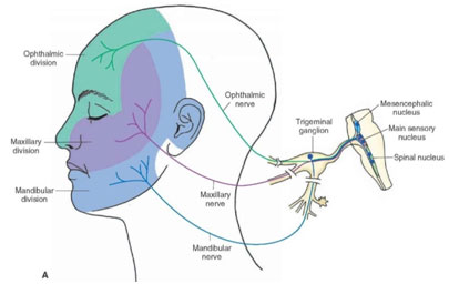
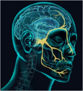 The trigeminal nerve is the fifth cranial nerve and is responsible for transmitting sensations from the face, brain lining (meninges), eyes, sinuses, TMJs, ears, salivary glands, mouth, nasal cavity and teeth to the brain. It is composed of three branches; the ophthalmic, maxillary, and mandibular (see above). It is also the nerve that controls the muscles used for chewing.
The trigeminal nerve is the main sensory cranial nerve protecting the most important structures underpinning our survival (brain, sight, smell, airway and eating). Thus any threat to this system, including pain, is physiologically wired for the patient to run away. It’s a miracle that any patient tolerates undergoing routine dental surgery whilst awake in the dental chair. Dentists are masters in managing anxious fearful patients during incredibly difficult procedures despite this instinctive enhanced reaction to pain in the trigeminal system.
Sensory dermatomes
Remember the skin over the parotid and lower ear lobe are supplied by C2 and C3 (not the trigeminal nerve).
Nucleii
The trigeminal nerve has multiple brainstem nuclei (sensory and motor) as well as many interconnections with other cranial nerves. It swaps parasympathetic fibers and taste fibers willy-nilly, and divides into dozens of terminal branches.

There are three sensory and one motor nuclei.
The sensory nuclei are arranged in a column which spans from the midbrain through the pons and medulla and into the upper cervical cord. The axons of these nuclei cross to the opposite side, ascending in the spinothalamic tract, to relay in the thalamic nuclei; from there, they end in the cerebral cortex. The sensory nucleus of CN V is connected to other motor nuclei of the pons and medulla. In addition, the descending sensory spinal tract receives somatic sensory fibers from CNs VII, IX, and X.
- mesencephalic nucleus: proprioreceptive fibers for muscles of the face,orbit, mastication, tongue. The proprioceptive fibers of CN V arise from the muscles of mastication and the extraocular muscles. They terminate in the mesencephalic nucleus. This nucleus has connections to the motor nucleus of CN V.
- main sensory nucleus: located in the upper pons, lateral to the motor nucleus is responsible for touch sensation for all three trigeminal divisions. The main sensory nucleus receives its afferents (as the sensory root) from the semilunar ganglion through the lateral part of the pons ventral surface. Its axons cross to the other side, ascending to the thalamic nuclei to relay in the postcentral cerebral cortex. The descending sensory fibers from the semilunar ganglion course through the pons and medulla in the spinal tract of CN V to end in the nuclei of this tract (as far as the second cervical segment).
The sensory nucleus, located in the pons, is quite extensive. It receives ordinary sensations from the main 3 branches of the trigeminal. The ophthalmic division is in the lower part of the nucleus, and the mandibular branch is in the upper part.
The large rostral head is the main sensory nucleus. The caudal tapered part is the spinal tract, which is continuous with substantia gelatinosa of Rolando in the spinal cord. The spinal tract is the sensory nucleus, primarily for pain and temperature. The main sensory nucleus serves mostly for discrimination sense.
- spinal nucleus: lower pons to upper cervical cord is responsible for pain and temperature; additionally it receives afferent fibers from the glossopharyngeal nerve and vagus nerve.
- Themotor nucleus is located in the upper pons and innervates the muscles of mastication as well as mylohyoid and tensor palati. The motor nucleus is ventromedial to the sensory nucleus. It lies near the lateral angle of the fourth ventricle in the rostral part of the pons. The mesencephalic nucleus is in the midbrain and receives proprioceptive fibers from all muscles of mastication.
The motor nucleus of CN V receives cortical fibers for voluntary control of the muscles of mastication. These fibers are mostly crossed. It also receives input from the mesencephalic and sensory nuclei. The axons emerge anterior to the sensory root from the lateral surface of the pons. This motor root joins the semilunar ganglion together with the sensory root.
Intracranial component
The trigeminal nerve exits at the mid pons into the prepontine cistern and passes anteriorly to Meckel cave where its fibers rely forming gasserian ganglion.
The semilunar (gasserian or trigeminal) ganglion is the great sensory ganglion of CN V. It contains the sensory cell bodies of the 3 branches of the trigeminal nerve (the ophthalmic, mandibular, and maxillary divisions). The ophthalmic and maxillary nerves are purely sensory. The mandibular nerve has sensory and motor functions.
The gasserian ganglion lies in a depression on the petrous apex, within a dural fold called the Meckel cave. The sensory roots of the 3 branches of CN V are received anteriorly. They then pass from the posterior aspect of the ganglion to the pons. The motor root passes under the ganglion to join the sensory division of the mandibular nerve and exits the skull through foramen ovale. The carotid plexus contributes sympathetic fibers to the gasserian ganglion.
Exracranial part
It then divides into three main branches:
Ophthalmic division (V1) please see flow chart summary of ophthalmic division courtesy Maliha Diwan
While passing at the lateral wall of the cavernous sinus inferior to trochlear nerve then through the superior orbital fissure the ophthalmic nerve divides into the frontal, lacrimal, and nasociliary nerves. The frontal nerve divides into the supraorbital and supratrochlear nerves. Nasociliary nerve branches include a communicating branch to the ciliary ganglion, short and long ciliary nerves, the posterior and anterior ethmoidal nerves (the latter divides into internal and external nasal branches), and the infratrochlear nerve.
Maxillary division (V2) please see flow chart summary of maxillary division courtesy Maliha Diwan
Passes in the lateral wall of cavernous sinus then through the foramen rotundum to exit skull. Then it crosses the pterygopalatine fossa and enters the orbit through theinferior orbital fissure and the infraorbital canal. It emerges through the infraorbital foramen to become the infraorbital nerve. Hence orbital blow out fracture is associated with loss of sensation over the maxilla.
Mandibular division (V3) please see flow chart summaries of mandibular sensory division and mandibular motor division courtesy Maliha Diwan
Passes through the foramen ovale and gives motor supply to the muscles of mastication.
Other Information
Mandibular (courtesy Maliha Diwan)
mandibular motor (courtesy Maliha Diwan)
Maxillary (courtesy Maliha Diwan)
opthalamic (courtesy Maliha Diwan)
Education menu

Physiotherapy for Orofacial Pain
A short introductory presentation by Dr Hedvig van der Meer who has recently joined our team:

Our guide to better sleep
By Georgina Gray Good quality sleep is one of our core health pillars. Not only is it vital for energy, mental health and hormone production, it is also important for disease prevention and weight control. We should be aiming for 7-9 hours of good quality sleep every...